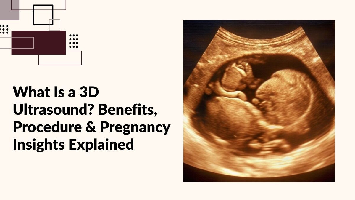
What Is a 3D Ultrasound? Benefits, Procedure & Pregnancy Insights Explained
Pregnancy is one of the most exciting journeys in life, and every parent looks forward to seeing their baby before birth. Thanks to advancements in medical imaging technology, a 3D Ultrasound allows parents and doctors to get a clearer and more detailed view of the developing baby. Unlike traditional ultrasound scans that produce flat, black-and-white images, a 3D Ultrasound Scan creates lifelike three-dimensional images, helping expectant parents visualize their baby’s facial features, movements, and development more clearly.
If you are searching for 3D Ultrasound in Jaipur, Ultrasound in Mansarovar, or looking for an Ultrasound near me, understanding how this advanced scan works can help you make informed decisions during your pregnancy.
What Is a 3D Ultrasound?
A 3D Ultrasound is an advanced imaging technique that uses sound waves to create three-dimensional images of the fetus inside the womb. The scan captures multiple two-dimensional images from different angles and combines them into a detailed 3D picture.
This technology has become increasingly popular because it provides enhanced visualization of the baby’s face, hands, feet, and overall physical development. A 3D Pregnancy Ultrasound is not only an emotional experience for parents but also an important diagnostic tool for healthcare professionals.
How Does a 3D Ultrasound Scan Work?
The procedure uses the same ultrasound technology as a traditional scan. A trained sonographer applies a special gel to the mother’s abdomen and moves a handheld device called a transducer over the skin.
The transducer sends high-frequency sound waves into the body. These sound waves bounce back from tissues and organs, creating images that are processed by advanced software into three-dimensional views.
The result is highly detailed Fetal Ultrasound Imaging that helps doctors assess fetal growth and detect certain developmental abnormalities more effectively.
Difference Between 2D and 3D Ultrasound
A 2D ultrasound and a 3D ultrasound both use sound waves to monitor fetal development during pregnancy, but they differ in the level of detail they provide. A traditional 2D ultrasound produces flat, black-and-white images that help doctors assess the baby’s growth, position, and overall health. In contrast, a 3D Ultrasound Scan creates detailed three-dimensional images, allowing clearer visualization of the baby’s facial features, hands, feet, and body contours. While 2D scans are commonly used for routine pregnancy assessments, 3D Sonography offers enhanced anatomical views that can help evaluate certain fetal structures more effectively. It also provides a more realistic and engaging experience for expectant parents, making it easier to connect with their unborn baby through lifelike images. Both scans play an important role in prenatal care, but a 3D Pregnancy Ultrasound delivers a more detailed and comprehensive view of fetal development.
While both scans are valuable, a 3D Sonography offers a more comprehensive visual experience.
Why Is a 3D Ultrasound Performed?
Doctors may recommend a 3D Ultrasound Scan for several reasons:
- Monitoring fetal development
- Evaluating facial structures
- Detecting cleft lip or palate
- Assessing fetal growth
- Examining limb development
- Monitoring high-risk pregnancies
- Providing detailed pregnancy imaging
In many cases, the scan offers additional information that may not be easily visible on standard ultrasound imaging.
Benefits of 3D Ultrasound During Pregnancy
1. Enhanced Visualization of Your Baby
One of the biggest advantages of a 3D Pregnancy Ultrasound is the ability to see your baby’s facial expressions and physical features more clearly. Parents often find this experience deeply reassuring and emotionally meaningful.
2. Better Detection of Certain Birth Defects
A 3D Ultrasound can help healthcare providers identify specific structural abnormalities such as:
- Cleft lip
- Cleft palate
- Facial abnormalities
- Limb deformities
- Neural tube defects
This detailed visualization allows for better pregnancy management and treatment planning when necessary.
3. Improved Bonding Experience
Seeing a realistic image of your unborn baby often strengthens the emotional connection between parents and their child. Many families cherish these images as memorable keepsakes.
4. Detailed Fetal Assessment
Advanced Fetal Ultrasound Imaging enables specialists to examine various parts of the baby’s anatomy with greater precision, helping ensure healthy fetal development.
5. Helpful in High-Risk Pregnancies
For pregnancies requiring closer monitoring, a 3D Ultrasound Scan can provide additional information that assists doctors in making informed clinical decisions.
When Is the Best Time to Get a 3D Ultrasound?
The ideal period for a 3D Ultrasound is generally between 24 and 32 weeks of pregnancy.
During this stage:
- The baby’s facial features are more developed.
- Adequate amniotic fluid surrounds the baby.
- Images tend to be clearer and more detailed.
However, the exact timing depends on the purpose of the scan and your doctor’s recommendations.
What Happens During the Procedure?
If you’re planning to undergo 3D Ultrasound in Jaipur, understanding the process can help ease any concerns.
Step 1: Preparation
You may be advised to stay hydrated before the scan to improve image quality. Wear comfortable clothing that allows easy access to your abdomen.
Step 2: Gel Application
A water-based gel is applied to the abdomen to help transmit sound waves effectively.
Step 3: Scanning Process
The sonographer gently moves the transducer over the abdomen while capturing multiple images from different angles.
Step 4: Image Processing
Advanced software combines these images into detailed three-dimensional views.
Step 5: Review and Discussion
The images are reviewed by a radiologist or fetal medicine specialist who interprets the results and discusses findings with you.
The entire process typically takes 20 to 45 minutes, depending on the baby’s position and the purpose of the examination.
Is 3D Ultrasound Safe?
Yes. A 3D Ultrasound Scan uses the same sound-wave technology as conventional ultrasound examinations.
It does not involve:
- Radiation exposure
- Surgical procedures
- Invasive instruments
When performed by qualified healthcare professionals, 3D Sonography is considered safe for both mother and baby. However, medical scans should always be conducted based on clinical need and professional guidance.
What Can You See in a 3D Ultrasound?
Depending on the baby’s position and gestational age, parents may be able to see:
- Facial features
- Nose and lips
- Fingers and toes
- Hands and feet
- Baby’s expressions
- Body contours
These realistic images provide a unique glimpse into your baby’s development before birth.
Choosing the Right Center for 3D Ultrasound in Jaipur
The quality of a Pregnancy Ultrasound Scan depends significantly on the expertise of the imaging center.
When selecting a diagnostic center, consider:
Experienced Specialists
Look for qualified radiologists and fetal medicine experts with extensive experience in pregnancy imaging.
Advanced Imaging Technology
Modern ultrasound systems produce clearer and more accurate images.
Comprehensive Pregnancy Care
A center offering routine scans, anomaly scans, growth scans, and advanced fetal assessments can provide better continuity of care.
Patient Comfort
A supportive environment helps expectant mothers feel comfortable throughout the procedure.
If you are looking for Ultrasound in Mansarovar or searching online for an Ultrasound near me, choosing a trusted diagnostic center with advanced imaging facilities can make a significant difference in both image quality and overall experience.
Frequently Asked Questions (FAQs)
Can a 3D Ultrasound Determine the Baby’s Gender?
In many cases, the baby’s gender may be visible during the scan. However, local regulations and medical guidelines must always be followed.
Does Every Pregnant Woman Need a 3D Ultrasound?
Not necessarily. Your doctor will recommend the most appropriate scans based on your pregnancy needs and medical history.
How Long Does a 3D Ultrasound Take?
Most scans take between 20 and 45 minutes, depending on fetal position and image quality.
What If the Baby Is Not Facing the Camera?
Sometimes the baby’s position may limit image clarity. Your sonographer may suggest changing positions or scheduling another appointment if necessary.
Can 3D Ultrasound Detect Birth Defects?
A 3D Ultrasound Scan can help identify certain structural abnormalities, but it is often used alongside standard pregnancy imaging and other diagnostic tests.
Final Thoughts
A 3D Ultrasound offers a remarkable opportunity to view your baby’s development in greater detail while providing valuable clinical insights for healthcare providers. From enhancing parental bonding to supporting detailed fetal assessments, this advanced imaging technology plays an important role in modern prenatal care.
Whether you’re considering a 3D Ultrasound in Jaipur, seeking reliable Ultrasound in Mansarovar, or searching for an Ultrasound near me, choosing an experienced diagnostic center with advanced imaging technology ensures accurate results and a comfortable experience throughout your pregnancy journey.
With proper medical guidance and timely scans, you can gain deeper insights into your baby’s growth while creating unforgettable memories during one of life’s most special moments.