What Are the Key Benefits of Ultrasound Imaging?
Ultrasound imaging is a widely used diagnostic technique that helps healthcare professionals examine structures inside the body without using ionizing radiation. It uses high-frequency sound waves to create real-time images of organs, tissues, blood vessels, and other internal structures. Because it is generally non-invasive, comfortable, and convenient, ultrasound has become an important part of modern diagnostic care.
People may be advised to undergo an ultrasound for several reasons, including evaluating abdominal organs, monitoring pregnancy, examining soft tissues, assessing blood flow, or investigating unexplained symptoms. Understanding its benefits can help patients know why doctors may recommend this imaging method.
What Is Ultrasound Imaging?
Ultrasound imaging, also known as sonography, uses a handheld device called a transducer. The transducer sends high-frequency sound waves into the body and receives the echoes that return from different tissues. A computer processes these echoes and converts them into images that can be viewed on a monitor.
Unlike imaging methods that use ionizing radiation, diagnostic ultrasound relies on sound waves. This makes it useful for a variety of examinations when a doctor needs to observe internal structures in real time.
Depending on the area being examined, different types of ultrasound examinations may be performed. These can include abdominal, pelvic, obstetric, breast, thyroid, vascular, musculoskeletal, and other specialized scans.
What Are the Key Benefits of Ultrasound Imaging?
1. It Does Not Use Ionizing Radiation
One of the commonly recognized advantages of diagnostic ultrasound is that it does not use X-rays or other forms of ionizing radiation. Instead, it uses high-frequency sound waves to produce images.
This characteristic makes ultrasound particularly useful when repeated imaging may be required. However, patients should still undergo an ultrasound when medically appropriate and follow their healthcare professional's recommendations.
2. Ultrasound Provides Real-Time Imaging
Ultrasound can display movement in real time. This allows healthcare professionals to observe organs and tissues while they are moving.
For example, ultrasound can help assess the movement of a fetus during pregnancy, observe blood flow using Doppler techniques, and evaluate certain soft-tissue structures. Real-time imaging can provide useful information that static images may not show as effectively.
3. It Is Generally Non-Invasive
Most ultrasound examinations are non-invasive. A healthcare professional usually applies a special gel to the skin and moves the transducer over the area being examined.
In many routine examinations, there is no need for an incision or injection. This can make the procedure more comfortable for patients and often allows the examination to be completed within a relatively short period.
4. It Can Examine Different Parts of the Body
Ultrasound is a versatile imaging technique that can be used to examine many areas of the body. Depending on the clinical requirement, it may be used to evaluate the liver, gallbladder, kidneys, pancreas, thyroid, uterus, ovaries, blood vessels, muscles, tendons, and other structures.
The specific type of scan depends on the patient's symptoms, medical history, and the information the doctor needs.
5. Ultrasound Can Support Early Medical Evaluation
When a patient experiences symptoms such as abdominal discomfort, swelling, unusual pain, or other concerns, a doctor may recommend imaging to investigate possible causes.
Ultrasound can provide visual information about certain organs and tissues, helping healthcare professionals evaluate abnormalities and decide whether additional tests may be required. It is important to remember that an ultrasound does not diagnose every medical condition on its own. Results should be interpreted along with symptoms, clinical examination, and other relevant investigations.
6. It Can Be Useful During Pregnancy
Ultrasound plays an important role in obstetric care. Healthcare professionals may use pregnancy ultrasound to monitor fetal development, assess growth, check fetal position, and evaluate various aspects of pregnancy.
Different pregnancy scans may be recommended at different stages depending on the pregnancy and clinical requirements. The timing and type of examination should always be determined by the treating healthcare professional.
7. Doppler Ultrasound Can Assess Blood Flow
Doppler ultrasound is a specialized form of ultrasound that can provide information about blood flow through blood vessels.
It may be used to assess the direction and movement of blood and can support the evaluation of certain vascular conditions. Doctors may recommend Doppler examinations when they need additional information about circulation in a particular area.
8. It Can Assist in Guiding Certain Procedures
Ultrasound can also be used to help healthcare professionals guide certain medical procedures. Real-time imaging can allow a clinician to identify the location of a structure while performing a procedure.
For example, ultrasound guidance may be used for selected needle-based procedures when clinically appropriate. The exact technique depends on the procedure, the patient's condition, and the healthcare professional's expertise.
When Might a Doctor Recommend an Ultrasound?
The reason for an ultrasound varies from person to person. A doctor may recommend an examination when they need more information about a particular symptom or suspected condition.
Common reasons can include:
- Investigating abdominal or pelvic symptoms
- Monitoring pregnancy
- Evaluating certain soft-tissue concerns
- Examining the thyroid or other superficial structures
- Assessing blood flow with Doppler imaging
- Evaluating certain lumps or swellings
- Monitoring specific medical conditions
- Supporting selected procedures
The doctor determines which type of ultrasound is appropriate based on the patient's individual clinical situation.
What Should You Know Before an Ultrasound?
Preparation depends on the type of ultrasound being performed. Some examinations may require the patient to drink water beforehand, while others may require fasting for a specific period.
For certain abdominal examinations, fasting may be recommended to improve visualization of particular organs. Pelvic examinations may sometimes require a full bladder.
Patients should follow the preparation instructions provided by the imaging center or healthcare professional. Following these instructions can help obtain clearer and more useful images.
If you are searching for Ultrasound in Mansarovar, check the imaging center's preparation instructions before visiting because requirements can differ depending on the examination being performed.
How to Find an Appropriate Ultrasound Facility?
Choosing an imaging facility involves more than simply looking for the closest option. Patients can consider factors such as the availability of the required scan, qualified healthcare professionals, appropriate imaging equipment, cleanliness, appointment availability, and accessibility.
People searching for Ultrasound in Jaipur may compare nearby diagnostic centers based on the type of examination they need. It can also be useful to confirm whether the facility provides specialized services such as Doppler, pregnancy ultrasound, abdominal ultrasound, or other examinations.
Patients should also take their doctor's prescription or recommendation with them when applicable so that the imaging team can understand the requested examination.
Is Ultrasound Safe?
Diagnostic ultrasound is generally considered a safe imaging technique when used appropriately for medical purposes. It does not involve ionizing radiation. However, as with any medical technology, it should be performed for an appropriate clinical reason and by trained professionals.
Patients should discuss any concerns with their doctor or the imaging professional before the examination.
What Happens During an Ultrasound Scan?
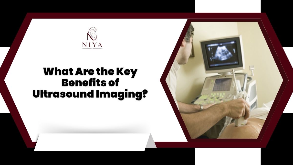
During a typical ultrasound examination, the patient may be asked to lie in a specific position depending on the body area being examined. A small amount of ultrasound gel is applied to the skin.
The sonographer or healthcare professional then moves the transducer over the examination area. The device sends sound waves into the body and receives returning echoes, which are converted into images.
The duration of the examination depends on the type of ultrasound and the area being evaluated. After the scan, the images are reviewed and a report may be prepared according to the facility's standard process.
What Are the Limitations of Ultrasound?
Although ultrasound offers many benefits, it is not suitable for every diagnostic situation. Sound waves do not travel equally well through all types of tissue. Air and bone can make it difficult to obtain clear images of certain structures.
Image quality can also depend on factors such as the body area being examined, the equipment used, and the patient's individual anatomy.
For this reason, a doctor may recommend another imaging method, such as CT or MRI, when more detailed information is required.
How Can You Find an Ultrasound Near You?
When looking for Ultrasound near me, consider the type of scan prescribed rather than choosing a facility based only on distance. Confirm that the center offers the required examination and ask about appointment availability and preparation requirements.
It is also helpful to check whether the imaging center has qualified professionals and appropriate equipment for the examination you need.
Conclusion
Ultrasound imaging offers several important benefits, including real-time imaging, non-invasive examination, versatility, and the absence of ionizing radiation. It can assist healthcare professionals in evaluating different organs and tissues, monitoring pregnancy, assessing blood flow, and investigating certain symptoms.
If your doctor has recommended an ultrasound, choose an appropriate diagnostic facility based on the required examination, professional expertise, equipment, preparation requirements, and accessibility. Most importantly, discuss your symptoms and concerns with a qualified healthcare professional so the right diagnostic approach can be selected for your individual needs.
Frequently Asked Questions About Ultrasound
What is the main purpose of an ultrasound?
The main purpose of an ultrasound is to create images of internal structures using high-frequency sound waves. Doctors can use these images to evaluate organs, tissues, blood vessels, and certain conditions.
Does an ultrasound use radiation?
No. Diagnostic ultrasound uses high-frequency sound waves rather than ionizing radiation.
How long does an ultrasound take?
The duration varies depending on the type of examination and the area being scanned. Some routine examinations can be completed relatively quickly, while specialized scans may take longer.
Do all ultrasound scans require preparation?
No. Preparation depends on the type of ultrasound. Some scans may require fasting or a full bladder, while others may not require special preparation.
Can ultrasound detect every health condition?
No. Ultrasound has limitations and cannot detect every medical condition. Depending on the symptoms and clinical requirement, a doctor may recommend additional tests or different imaging methods.